Portfolio
A collection of scientific illustrations (and an animation) that I produced during my PhD work. Click on the images to enlarge them.
A collection of scientific illustrations (and an animation) that I produced during my PhD work. Click on the images to enlarge them.
A short animation showing how the gene expression of Hes5 in the developing mouse neural tube evolves over time and the migration of cells as they differentiate. There is an interactive simulation based on this tissue that you can find here.
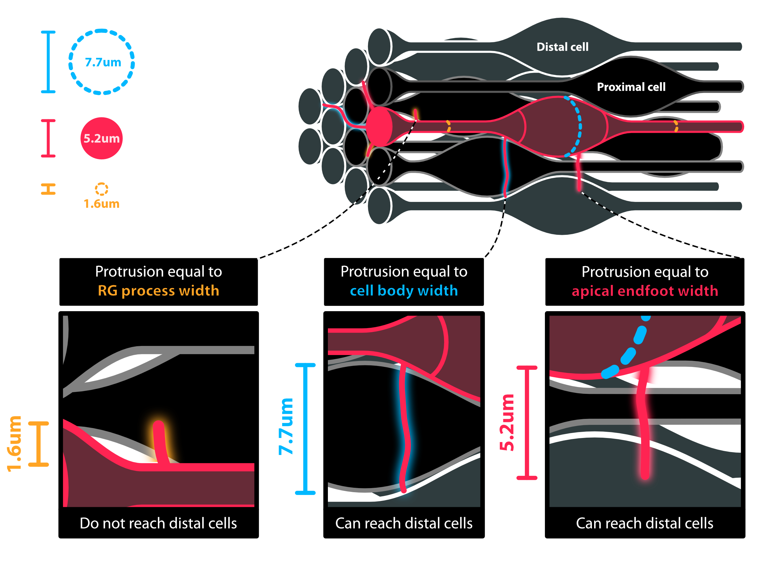
Idealised illustration of the structure of mouse neuroepithelial cells and how membrane protrusions relate to the experimentally measured cell widths.
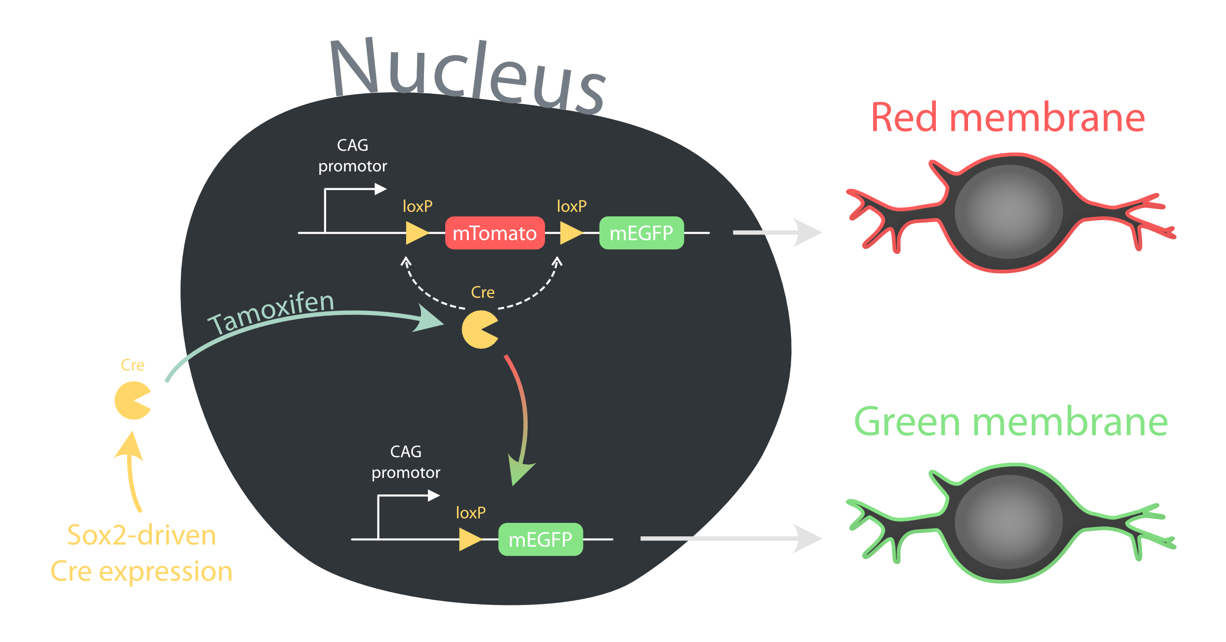
Summary of the Sox2CreERT2 mTmG system used to mosaically label the membranes of neural progenitor cells in the neural tube.
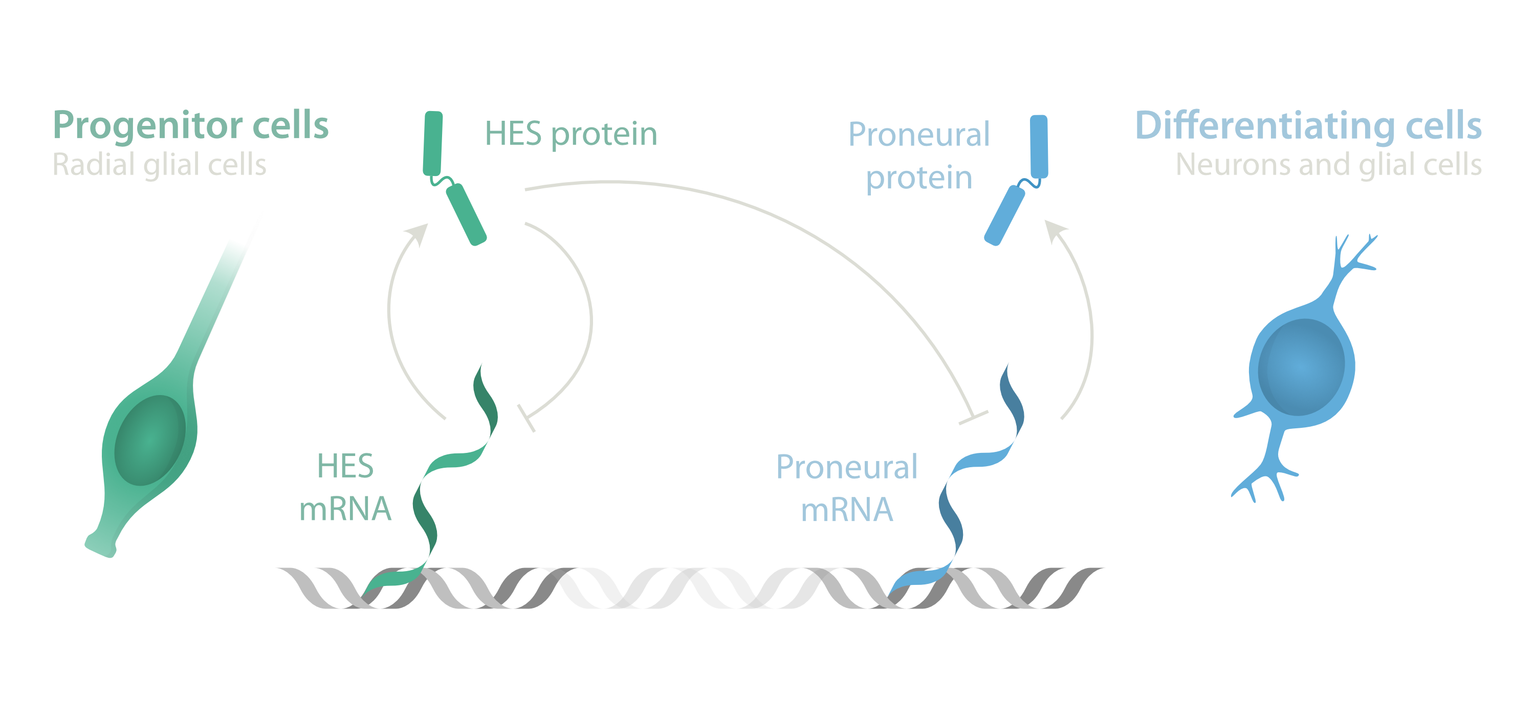
An overview of Hes5 gene regulatory interactions and the corresponding cell-states. Used in seminar talks.
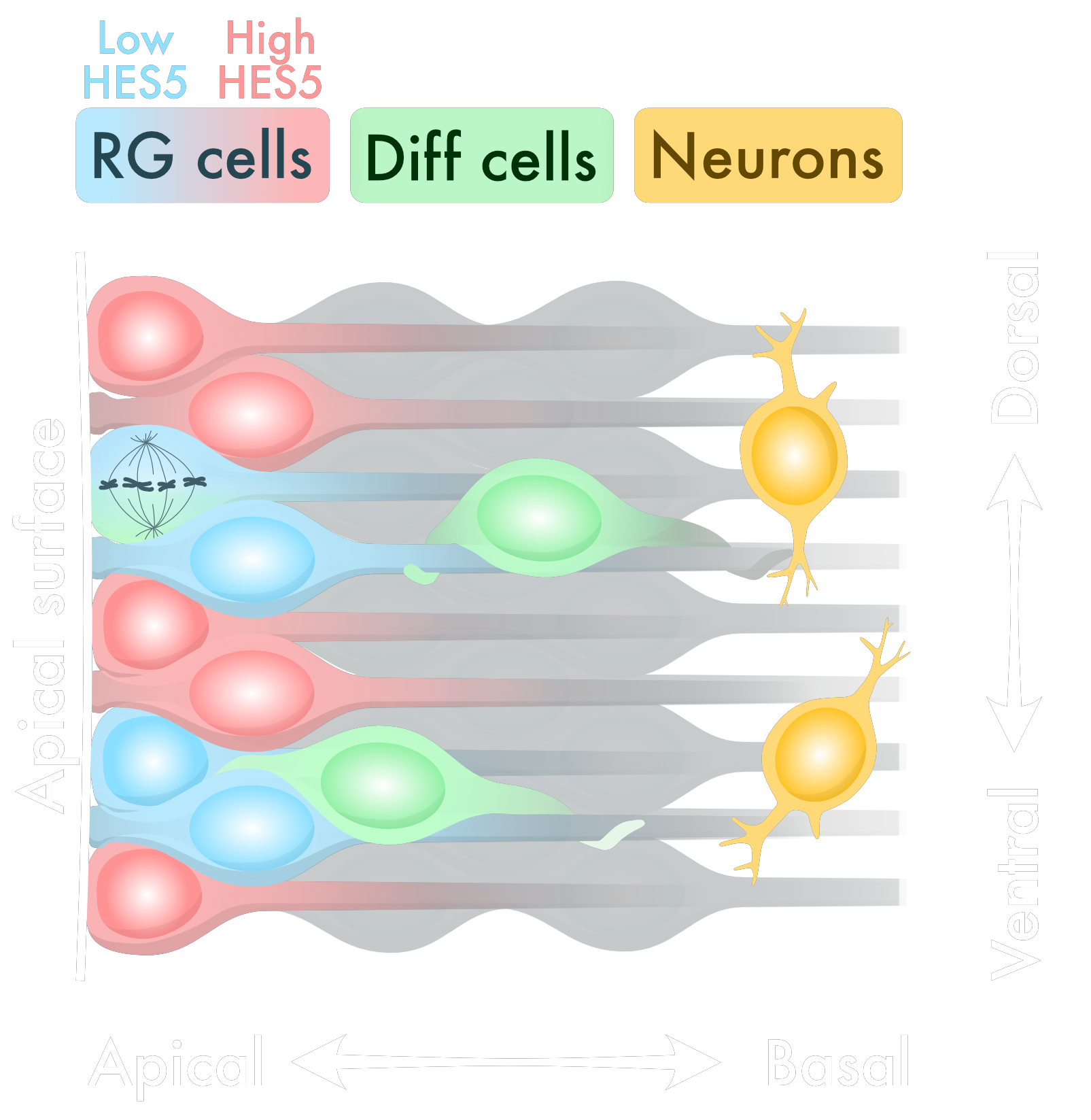
Part of a figure from my most recent paper, specifically this shows the cellular structure of the neural tube.

Illustration from a figure in my most recent paper, showing how a simulated grid of cells maps onto a biological structure called the neural tube.
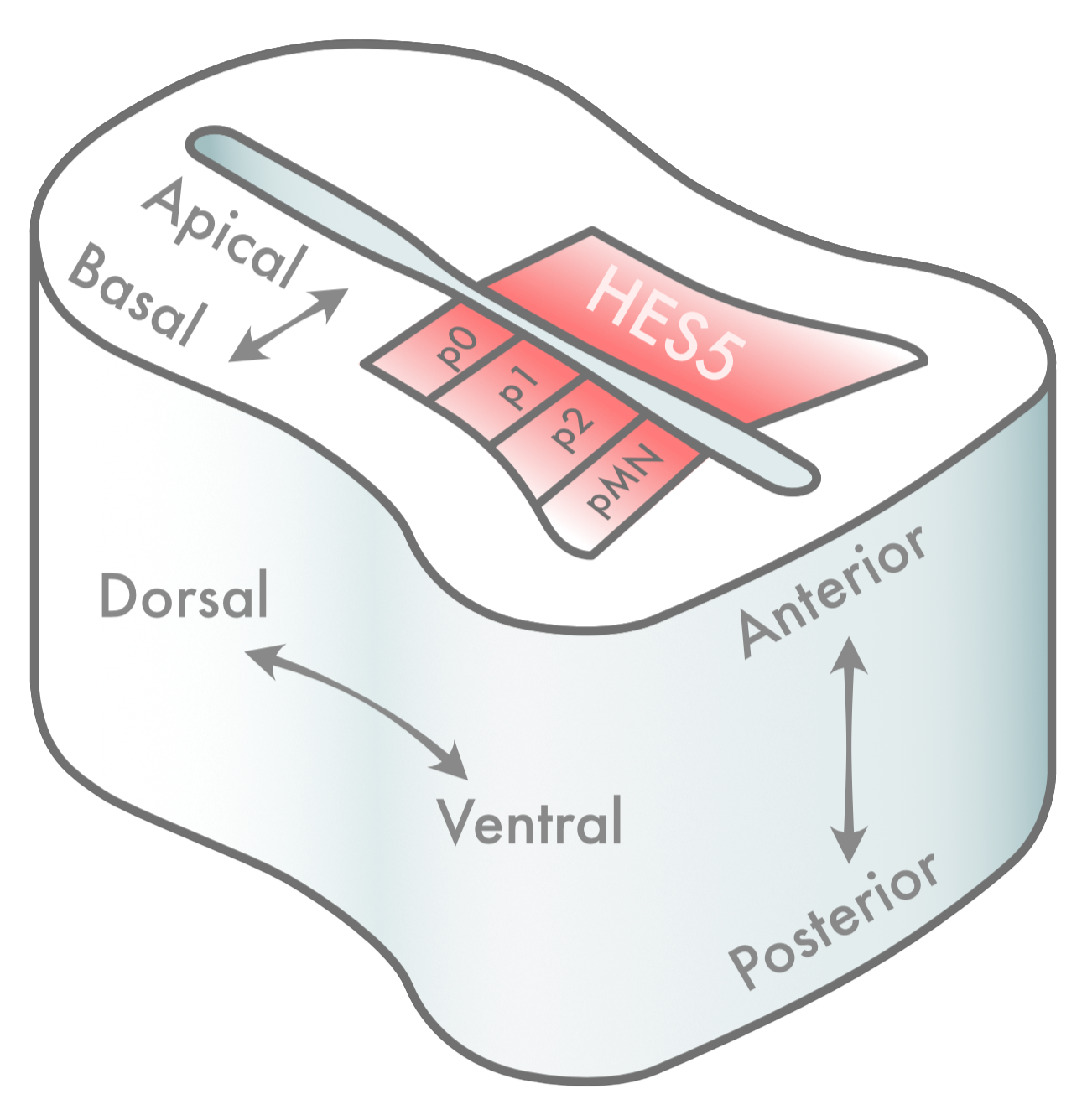
Where Hes5 is expressed within the developing neural tube. Taken from a figure in my most recent paper.

A graphical abstract that I produced for a colleague's recent paper.
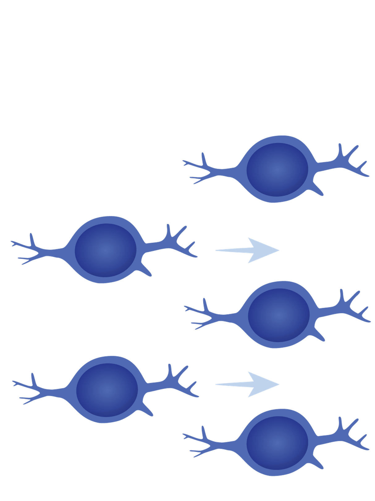
Seminar illustration of a gap-filling hypothesis mechanism that might operate during differentiation of neural stem cells in the developing spinal cord.